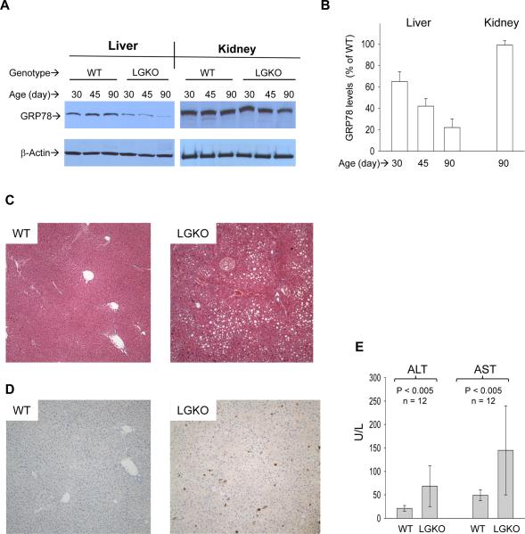
Figure 1. Liver specific loss of GRP78/BiP and liver injury.
(A) Western blots showing liver specific loss of GRP78 protein. WT, wild type littermates; LGKO, carrying the homozygous Grp78 floxed alleles and half copies of the cre transgene. (B) Graph depicts % of GRP78 of LGKO compared to that of WT. (C) H&E staining (×100). Showing fat accumulation in LGKO. (D) Hepatic apoptosis (brown spots) revealed by immunohistochemistry (100×) with anti-active caspase 3 antibodies. (E) Serum alanine aminotransferase (ALT) and aspartate aminotransferase (AST). Values are mean ± sd.

