
Figure 4.
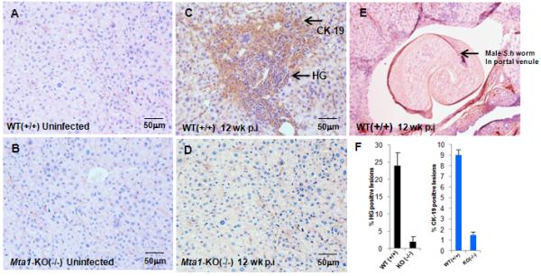
Influence of MTA-1 on formation of granulomatous inflammatory lesions in liver. Paraffin embedded liver tissue sections from age-matched WT and Mta1−/− mice at 5 and 12 weeks after exposure to Schistosoma haematobium cercariae were stained with anti-cytokeratin-19 (Ck-19) antibody followed by hematoxylin and eosin (H & E). Stained slides were scored under phase contrast microscopy for hepatic granulomatous (HG) regions. Representative liver sections from uninfected, control mice (panel A, WT Mta1, panel B, Mta1−/−), infected mice at 12 weeks (C, D) after infection. Regions showing high infiltration of polymorphonuclear cells were scored as positive for inflammation. Percentage HG positive and CK19 positive zones from 20 fields were determined and represented as bar plot (panel F). Panel E shows an adult male S. haematobium in situ in a portal venule in a WT mouse 12 weeks after exposure to cercariae (H&E stain). Images were captured under a 20 × objective.