
Figure 5.
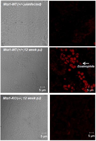
Mta1-WT mice show a higher degree of eosinophil infiltration in liver compared to age matched Mta1(−/−) mice 12 weeks post-infection. Liver tissue sections from infected WT and Mta1(−/−) mice were stained for eosinophils using EO-probe kit. Sections were scanned using Zeiss 710 confocal microscope and images were recorded using a 40× objective.