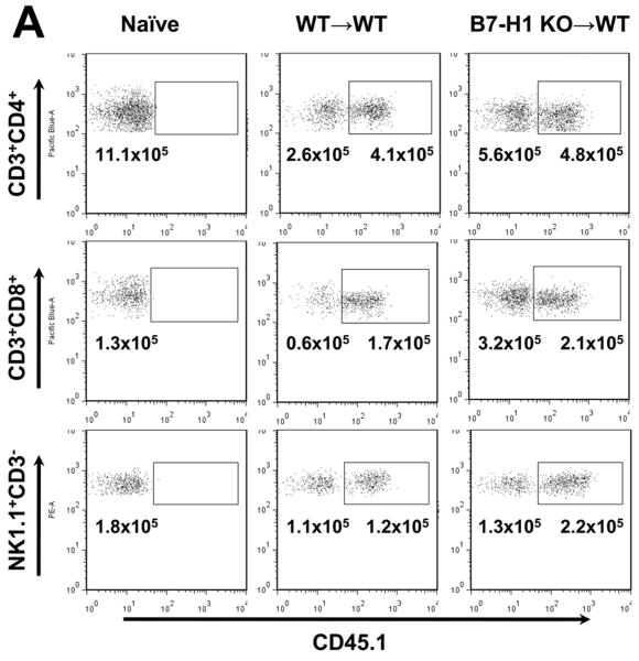
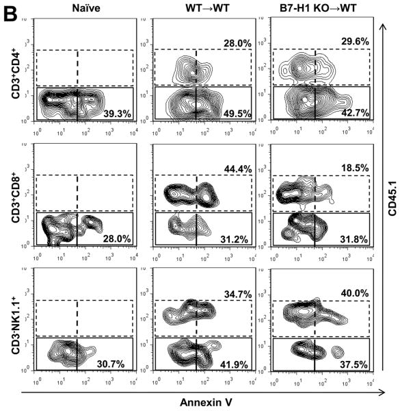
Figure 4. Analysis of graft- and host-derived CD4 T cells, CD8 T cells and NK cells.
(A) Flow cytometric analysis of liver NPC for donor/recipient phenotype. Hepatic NPC from B7-H1 KO and WT grafts 6 hr after LTx into CD45.1 recipients were analyzed. CD3+CD4+, CD3+CD8+ and NK1.1+CD3- cells were analyzed for CD45.1 (recipient) expression. Numbers are absolute numbers in liver grafts. Data are representative of three independent experiments.
(B) T cell apoptosis was determined by flow cytometry in liver NPC. CD3+CD4+, CD3+CD8+ and NK1.1+CD3- cells were analyzed for CD45.1 and Annexin V expression. Annexin V expression by host CD45.1+ CD8, but not CD4 or NK cells was reduced in KO grafts compared with WT grafts. No differences were detected in Annexin V expression by graft CD45.1- CD4, CD8 or NK cells between KO and WT grafts. Data are representative of three independent experiments.


