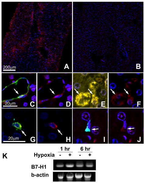
Figure 8. B7-H1 expression in human liver graft biopsy samples and cultured hepatocytes.
Liver graft biopsy samples (n=3 for normal, n=16 for post-reperfusion) were analyzed by multiplex quantum dot (QD) staining using antibody combinations of B7-H1 (red)/CD31 (endothelial cells, green)/CK19 (biliary epithelial cells, cyan)/HepPar1 (hepatocytes, yellow) or B7-H1 (red)/CD11c (DC, green)/CD68 (Kupffer cells, cyan). Representative images were selected.
(A) B7-H1 expression (red) was observed in perivenular lobular areas of post-reperfusion biopsy. (B) In contrast, normal liver samples showed only scarce B7-H1 expression largely limited to endothelial cells. (C, D) In the post-reperfusion biopsy, CD31+ endothelial cells (C, green) in the dilated sinusoids expressed B7-H1 (D, red). (E, F) HepPar1+ hepatocytes (E, yellow) in post-reperfusion biopsy specimens also showed B7-H1 expression (F, red). (G, H) CD11c+ CD68- DC (G, green) showed B7-H1 (H, red). (I, J) CD68+ CD11c- Kupffer cells (I, cyan) also showed B7-H1 expression (J, red).
(K) Primary human hepatocytes showed mRNA upregulation for B7-H1 when exposed to hypoxia. Representative image of two similar experiments.

