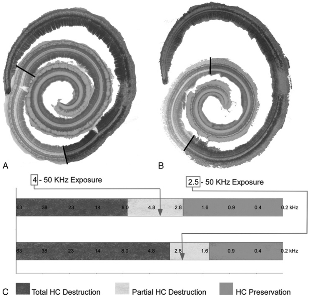
FIG. 3.
Histologic replication of residual hearing in the gerbil model. A, Color-coded representation of the extent of the 3 regions defined in the cochlea of Figure 2. B, Specimen from a separate case with a high-pass cutoff of 2.5 kHz (Gerbil 70). C, The degree of hair cell damage from each case with superimposed tonotopic frequency information. As anticipated, a larger region of complete hair cell loss (red area) is observed with the wider (2.5 kHz cutoff) noise exposure. The arrows mark the cutoff frequencies for each case, both of which fall within the zone of partial HC destruction.

