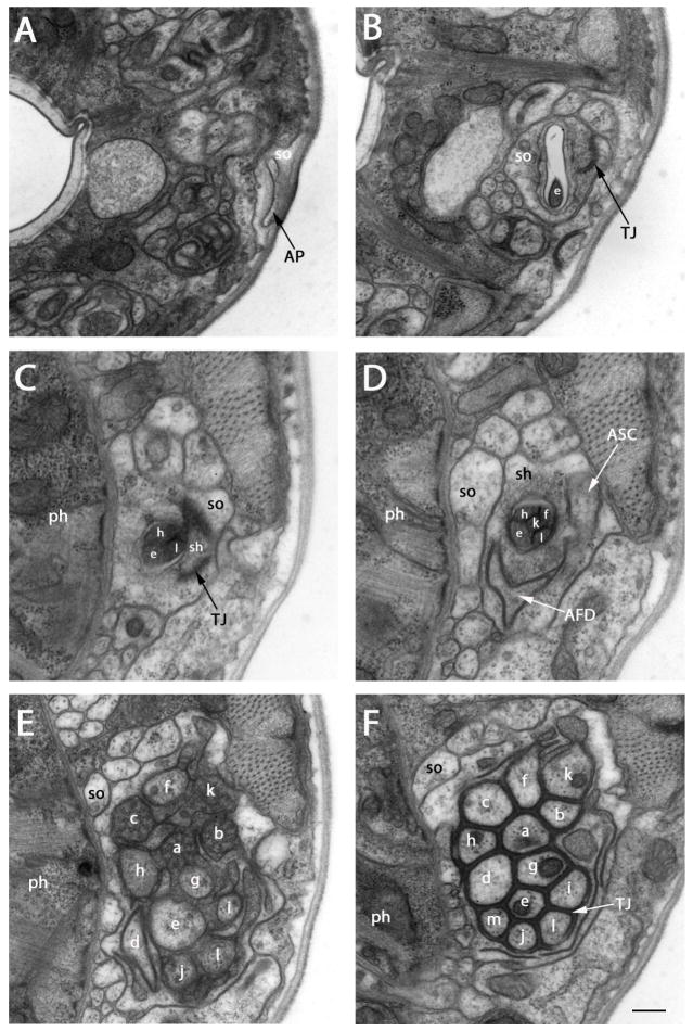
Figure 1.
Transmission electron micrographs of lateral sections at several levels between amphidial pores and the tight junction region of P. trichosuri. The sensory dendrites of ASA (a), ASB (b), ASC (c), AFD (d), ASE (e), ASF (f), ASG (g), ASH (h), ASI (i), ASJ (j), ASK (k), ASL (l) and ASM (m) are shown. A: Section approximately 2 μm posterior to the cephalic extremity. The amphidial pore (AP), which is formed by the socket cell (so) and communicates with the external environment is seen. B: Section approximately 1.5 μm posterior to the amphidial pore. At this level the cross section of ASE is found within the amphidial channel formed by the socket cell (so). TJ indicates the self-tight junction of the socket cell. C: Section approximately 4.5 μm posterior to the amphidial pore. Cross sections of three neurons, ASE, ASH and ASL, are found within the amphidial channel formed by the sheath cell (sh; ph=pharynx). This level contains the transition zone between the socket cell and the sheath cell. (TJ) (see above) indicates the tight junction between these two cells. D: Section approximately 5 μm posterior to the amphidial pore. Cross sections of five neurons, ASE, ASF, ASH, ASK and ASL, are found within the amphidial channel at this level. The finger-like projection (white arrow) of the finger cell (AFD) and the single dendrite of ASC (white arrow) are contained within the sheath cell. E: Section approximately 9.5 μm posterior to the amphidial pore. This level shows the transition zones of twelve neurons. F: Section approximately 10.2 μm posterior to the amphidial pore. At this level tight junctions (TJ) form between all 13 neurons and between the neurons, the sheath cell and the shortest neuron (ASM). Scale bar in panel F=2 microns and applies to all panels.

