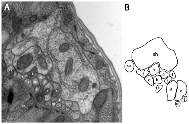
Figure 2.
Section approximately 11.2 μm posterior to the amphidial pore (1μm posterior to the tight junction region). A: After the tight junction region, which is at the posterior extremity of the amphidial channel, dendrites of all amphidial neurons, the socket cell and the sheath cell form a bundle and extend posteriorly to the lateral ganglion. B: Diagram identifies the neurons individually. Scale bar = 2 microns. Panel A corresponds to the plane labeled Fig. 2A in the three-dimensional reconstruction, Fig. 6B.

