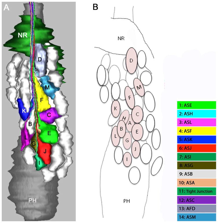
Figure 7.
Three-dimensional reconstruction of the amphidial cell bodies of Parastrongyloides trichosuri. Anterior is up. A: Model of the lateral ganglion to the right of the pharynx from a lateral view. The colored cell bodies are those of the amphidial neurons. Cell bodies of other unidentified neurons are represented in white. B: Diagram identifies the neuronal cell bodies individually. Amphidial cell bodies are tinted and marked with letters corresponding to the third letter of their unique three-letter designations. Unidentified cell bodies are represented as untinted ovals.

