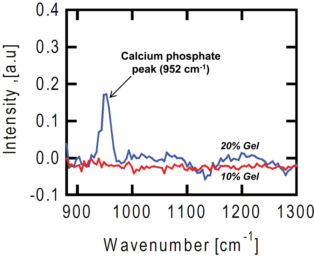
Figure 8.
Coherent anti-Stokes Raman scattering (CARS) imaging was used to analyze the composition of mineral deposits in gels. Osteoblasts encapsulated in control gels of uniform composition (10 % PEGDM, red line; 20 % PEGDM, blue line) were cultured for 21 d, fixed and imaged by CARS. The softer gels (10 % PEGDM, 46 kPa) did not mineralize whereas the stiffer gels (20 % PEGDM, 390 kPa) did mineralize, similar to the images shown in Fig. 7a where 20 % gels containing cells turned white. The peak at 952 cm−1 in the stiffer 20% gels (blue line) corresponds to a vibrational resonance for calcium phosphate, which is not observed in the spectra from the softer 10% gels (red line).

