
Figure 6. Interaction of Hsp20 with PP1.
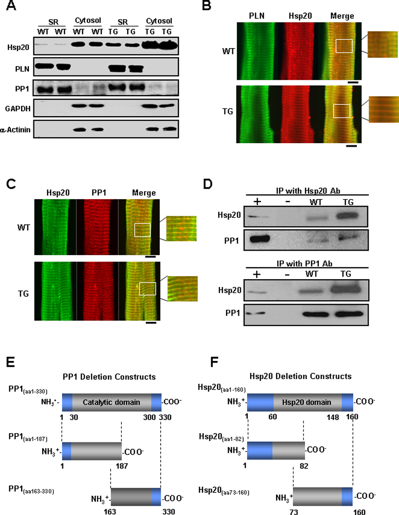
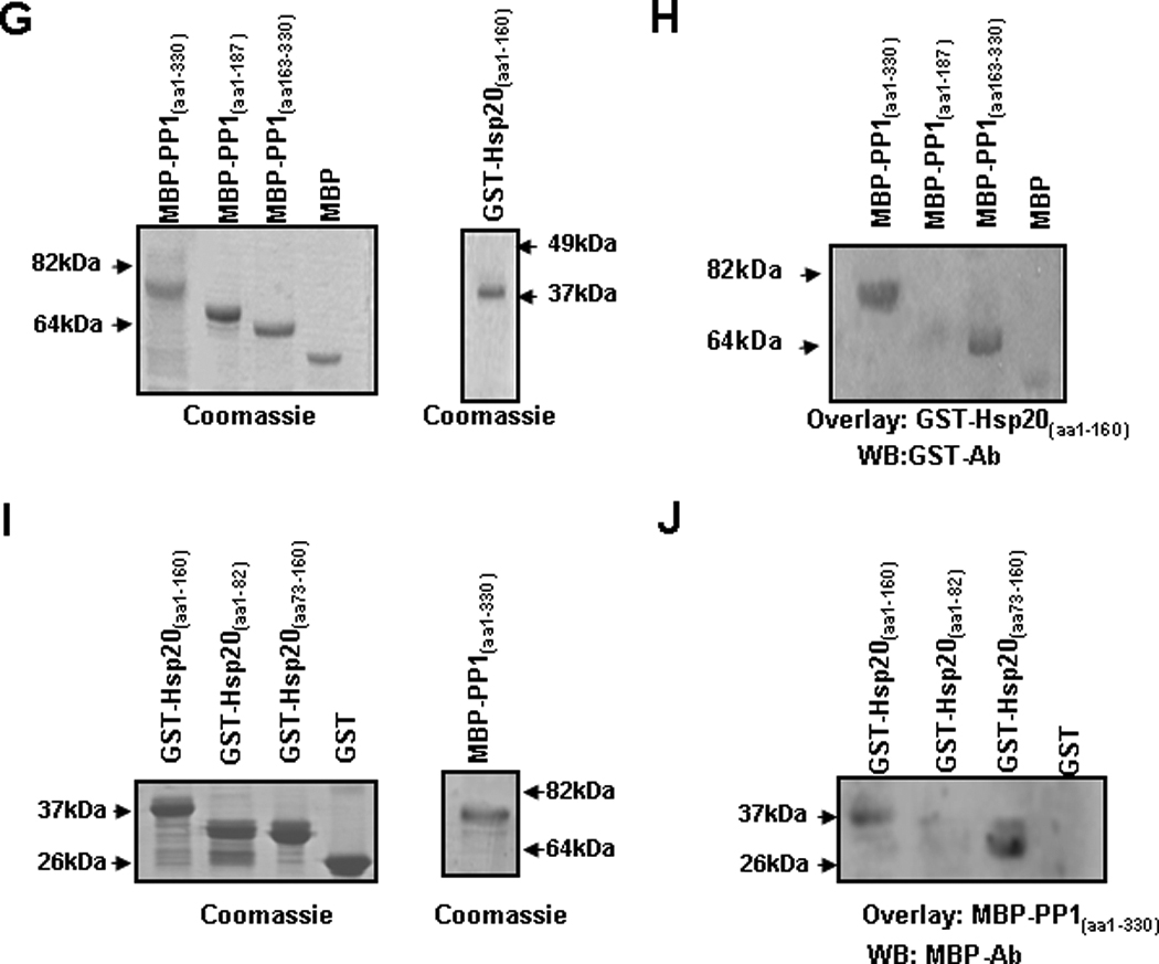
(A) Representative blots of Hsp20, PLN, PP1, GAPDH and α-actinin in microsomes enriched in SR membranes or in cytosolic fractions from WT and Hsp20 TG hearts (n=6). The yield of cardiac SR and cytosolic proteins averaged 1.5mg/heart and 12mg/ heart, respectively. The amount of total protein on each gel lane was the same (20 µg for each fraction), which diluted the cytosolic fraction by 10× compared to the SR fraction. (B) WT and TG cardiomyocytes were immunostained for Hsp20 (red) in combination with PLN (green). (C) WT and TG cardiomyocytes were immunostained for Hsp20 (green) in combination with PP1 (red). Scale bar, 10 µm. (D) Co-immunoprecipitation was performed using anti-Hsp20 or anti-PP1 antibody and cardiac homogenates (200µg total protein) of wild type and Hsp20 TG mice. The precipitates were analyzed by immunoblotting with anti-Hsp20 or anti-PP1 antibodies, as indicated. Preimmunoprecipitated WT heart homogenate was used as positive control (+), and immunoprecipitate with anti-IgG PLUS agarose was used as negative control (−). IP: immunoprecipitation. (E) Diagrammatic representation of the full length and the two deletion constructs of PP1 and (F) the full length and the two deletion constructs of Hsp20. Predicted protein domains are shown in grey. (G) SDS-gel stained with Coomassie blue showing the purified MBP-PP1 full-length or deletion proteins. (H) Blot overlay assays with anti-GST antibody (WB : GST-Ab) were performed to determine the protein region of PP1 required for its association with GST-Hsp20(aa1–160). This narrowed down the PP1 binding region to a C-terminal fragment including amino acids 163–330. (I) SDS-gel stained with Coomassie blue showing the purified GST-Hsp20 full-length or deletion proteins. (J) Blot overlay assays with anti-MBP antibody (WB: MBP-Ab) determined that the protein region of Hsp20 responsible for its binding with MBP-PP1(aa1–330) includes amino acids 73–160. WB: Western blot.