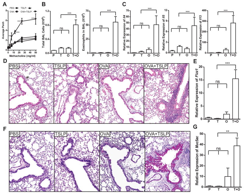
Figure 1. TSLP diverts airway tolerance to Th2 sensitization against innocuous antigen OVA.
BALB/c mice were conditioned i.n. with PBS, TSLP, OVA, or OVA + TSLP for 3 days. Ten days later, the animals were challenged i.n. with OVA for three times. A, Airway responsiveness to increasing dose of methacholine was analyzed by unrestrained whole body plethysmography and is presented as average enhanced pause (Penh) over a three minute period. B, Total cell and eosinophil count in BAL fluid. C, Quantitative PCR analysis of the expression of Th2 cytokines Il-4, Il-5 and Il-13 in the lungs, presented relative to the expression in PBS conditioned and OVA challenged mice. D, Lung tissue sections stained with H&E showing peribronchiolar inflammatory infiltration in OVA + TSLP conditioned mice after challenge. E, Quantitative PCR analysis of the expression of Fizz1 gene in the lungs, presented relative to the expression in PBS conditioned and OVA challenged mice. F, Lung tissue sections stained with PAS showing goblet cell hyperplasia/metaplasia in OVA + TSLP conditioned mice after challenge. G, Quantitative PCR analysis of the expression of mucus gene Muc5ac, presented relative to PBS conditioned and OVA challenged mice. P: PBS-conditioned mice; T: TSLP-conditioned mice; O: OVA-conditioned mice; T + O: OVA plus TSLP conditioned mice. Data shown represent mean ± SEM (n = 4) from one of two independent experiments. **: p < 0.01; ***: P < 0.001; ns: not significant by analysis of variance with Bonferroni’s post-hoc tests.

