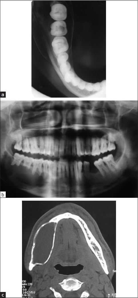
Figure 1.

(a) Mandibular occlusal radiograph showing expansion of the cortical plates; (b) panoramic radiography showing a unilocular radiolucency extending from 45 to 48 region; (c) axial CT image of the mandible showing cortical expansion and thinning
