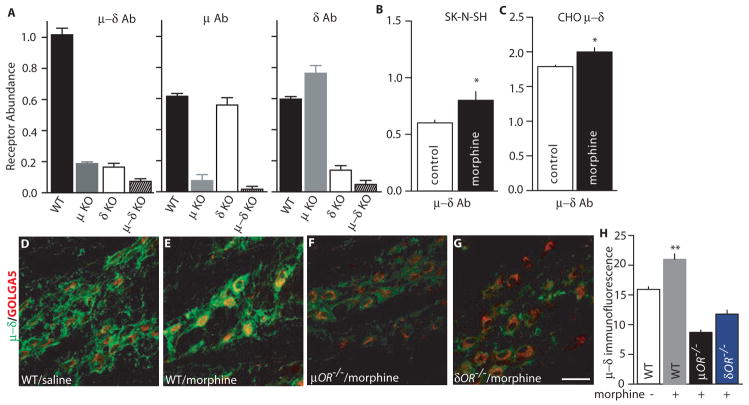
Fig. 1.
Detection of μ–δ heteromers using heteromer-selective monoclonal antibodies. (A) Receptor abundance was determined in cortical membranes from wild-type, μ knockout (KO), δ KO, or μ–δ double KO mice with monoclonal antibodies to μ–δ, μ, or δ receptors by ELISA. (B–C) Cells endogenously (B) or stably (C) coexpressing μ–δ receptors were treated without or with morphine (1 μM) for 48 hours. Heteromer abundance was determined by ELISA with μ–δ heteromer-selective antibodies. Results are means ± SEM (n = 3 experiments). *p < 0.05. (D–G) Repeated morphine treatment increased μ–δ heteromer immunoreactivity in the medial nucleus of the trapezoid body (MNTB). Immunoreactivity in μ KO or δ KO mice after morphine treatment was below the detection limits. (H) Individual neuronal profiles were outlined (n = 15–20/group) and μ–δ heteromer immunoreactivity, as assessed by mean optical density (O.D.), was determined. Morphine treatment increased μ–δ heteromer immunoreactivity in MNTB neurons from wild-type mice (p < 0.01 compared to untreated control), but not in those from μ or δ KO mice.

