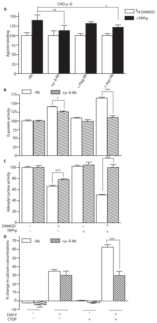
Fig.4.
μ–δ heteromer-selective antibody blocks heteromer-mediated increases in binding and signaling. (A) CHO cells coexpressing Flag-tagged μ and Myc-tagged δ receptors were pre-treated with either the μ–δ heteromer-selective antibody or monoclonal antibodies to Flag or Myc. Binding of the μ receptor agonist [3H]DAMGO (10 nM) to cells was measured in the absence or presence of δ receptor antagonist TIPPψ (10 nM). Results are means ± SEM (n=3 experiments). (B and C) Mouse cortical membranes were preincubated without or with μ–δ heteromer antibodies and G-protein activity, as assessed by [35S]GTPγS binding (expressed as G-protein activity), (B) or adenylyl cyclase activity (C) in response to DAMGO (1 μM) was determined in the absence or presence of TIPPψ (10 nM). Results are means ± SEM (n=3 experiments). (D) HEK293 cells coexpressing chimeric G16/Gi3 and μ and δ opioid receptors were preincubated without or with μ–δ heteromer antibodies, then treated with the δ opioid agonist deltorphin II (1 μM) in the absence or presence of the μ opioid antagonist CTOP (10 nM) and intracellular Ca2+ concentrations were determined. Results are means ± SEM (n=3 experiments). *p<0.05; **p<0.01; ***p<0.001

