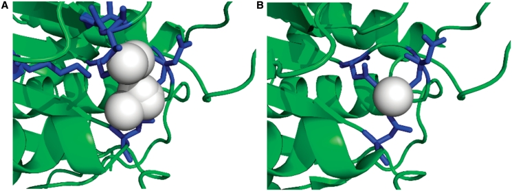
Figure 4.
Predicted and observed binding site residues and ligands, for CASP9 target T0635 (PDBID 3n1u). (A) The FunFOLD 1.0 results with the top model magnified to zoom in on the binding site, the binding site residues are colored blue (25,27,69,70,95,118) and the predicted ligand cluster, with frequencies of putative ligands, colored in white (CL-2, CA-3, SO4-2, PO4-1, MG-5, CO-1). (B) The observed binding site for the native hydrolase (PDBID 3n1u and CASP9 target T0635), with the binding site residues colored blue (25, 27, 118) and the observed ligand (CA) colored white. The binding site prediction has an MCC score (16) of 0.7012 and BDT score (17) of 0.5744.

