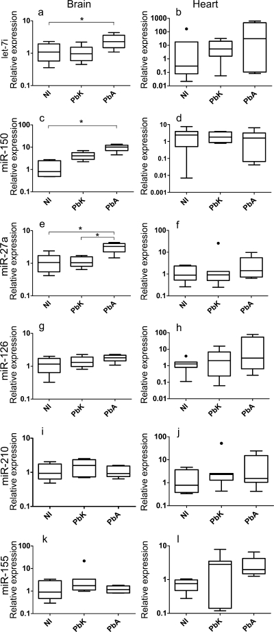
Fig. 1.
Inflammation-associated miRNAs in brain and heart tissues in cases of cerebral (PbA) versus noncerebral (PbK) malaria and NI tissues. Box plots show miRNA expression levels measured for NI and PbK- and PbA-infected brain and heart tissues, expressed as normalized values using miR-U6 as the endogenous control. The horizontal line denotes the median value, the box encompasses the upper and lower quartiles, whiskers show 1.5+ the interquartile range (Tukey), and outliers are denoted with a closed circle. We detected a significant increase in the expression of let-7i (P value of 0.017), miR-27a (0.006), and miR-150 (<0.001) in brain tissue following PbA infection. If the Kruskal-Wallis test was significant, post hoc tests were carried out. The results of these are denoted in the plot with horizontal bars and asterisks (*, P < 0.05). No changes were discernible in miR-126 (P = 0.16), miR-155 (P = 0.31), and miR-210 (P = 0.34) following infection. No changes in expression were detected in heart tissue. These data represent results of three independent experiments, with 7 animals in each group.

