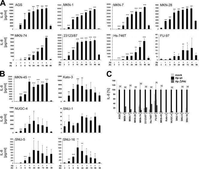
Fig. 6.
IL-8 secretion by H. pylori-infected gastric epithelial cells. (A and B) All adherent cells (A) and cells in suspension (B) were either left untreated (−) or infected with H. pylori at an MOI of 50 for 96 h. IL-8 secretion was determined by ELISAs, comparing each time point with its corresponding noninfected control. Only the controls of noninfected cells after 3 h and 96 h are shown. Data are presented as the mean values with standard errors from four independent experiments with three replications. Asterisks indicate statistically significant differences between H. pylori-induced IL-8 secretion after a given time point and its corresponding mock-infected control (*, P < 0.05; **, P < 0.01; ***, P < 0.001). (C) Cells were infected with wild-type H. pylori or its isogenic ΔPAI mutant at an MOI of 50 for 24 h or were left untreated. Aliquots of supernatants were analyzed for IL-8 secretion. The amounts of IL-8 after wild-type H. pylori infection were set 100%. Data are presented as the mean values with standard errors from two independent experiments with three replications. Asterisks indicate statistically significant differences between wild-type and ΔPAI H. pylori-induced IL-8 secretion (*, P < 0.05; **, P < 0.01; ***, P < 0.001).

