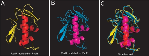
Fig. 6.
Models of the C-terminal domain of RevR. (A and B) The RevR amino acid sequence was modeled on the crystal structure of the PhoB effector domain from E. coli (Protein Data Bank [PDB] template code 1GXQ) (A) or the YycF effector domain from B subtilis (PDB template code 2D1V) (B) using the SWISS-MODEL server (http://swissmodel.expasy.org/). (C) The two hypothetical RevR models are superimposed.

