Fig. 3.
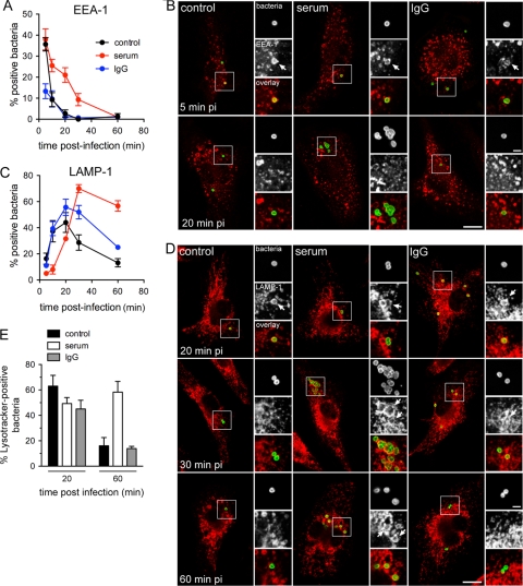
Serum opsonization of Schu S4 alters FCP early trafficking. BMMs were infected with either unopsonized (control), serum-opsonized (serum), or IgG-opsonized (IgG) Schu S4. At various times postinfection, samples were fixed and processed for immunolabeling of bacteria and either EEA-1 or LAMP-1. (A) Quantification of colocalization of intracellular Schu S4 with EEA-1. Data are means ± standard deviations of three independent experiments. (B) Representative confocal micrographs of BMMs that were infected with either unopsonized (control), serum-opsonized (serum), or IgG-opsonized (IgG) Schu S4 for either 5 or 20 min. Bacteria (appearing in green) are within EEA-1-positive FCPs (appearing in red) at 5 min p.i. Arrows in insets indicate bacteria within EEA-1-positive FCPs. Bars, 10 or 2 (inset) μm. (C) Quantification of colocalization of intracellular Schu S4 with LAMP-1. Data are means ± standard deviations of three independent experiments. (D) Representative confocal micrographs of BMMs that were infected with either unopsonized (control), serum-opsonized (serum), or IgG-opsonized (IgG) Schu S4 for either 20, 30, or 60 min. Arrows in insets indicate bacteria (appearing in green) within LAMP-1-positive FCPs (appearing in red). Bars, 10 or 2 (inset) μm. (E) Quantification of colocalization of intracellular Schu S4 with the acidic probe Lysotracker DND-99. BMMs were infected with either unopsonized (control), serum-opsonized (serum), or IgG-opsonized (IgG) GFP-expressing Schu S4; loaded with Lysotracker DND-99, as described in Materials and Methods; and analyzed at 20 and 60 min p.i. Values are means ± standard deviations of three independent experiments.

