Fig. 1.
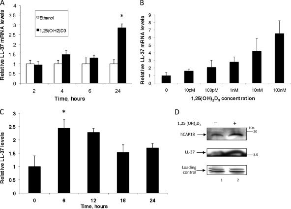
Induction of LL-37 in gingival epithelial cells in response to 1,25(OH)2D3. OKF6/TERT (A and B) or primary gingival epithelial cells (C) were cultured in the presence of 10−8 M 1,25(OH)2D3 or ethanol (0.1%) for increasing times (A and C) or for 24 h in the presence of increasing concentrations of 1,25(OH)2D3 (B). Total mRNA was isolated, and LL-37 mRNA levels were quantified by QRT-PCR. Results are mRNA levels of 1,25(OH)2D3-stimulated cultures compared to those of ethanol-treated cultures at each time point, normalized to the level for β2M (n = 3; bars indicate mean ± standard deviation). (D) LL-37 protein levels were visualized by Western blot analysis of whole-cell lysates of OKF6/TERT cells cultured in an air-liquid interface (ALI) and stimulated with 1,25(OH)2D3 for 18 h. Tubulin was visualized as a loading control. The increase in LL-37 mRNA levels in panel A is significant at 24 h as quantified by t test (P < 0.001). The dose response in panel B is significant as measured by analysis of variance (ANOVA) (P < 0.0001).

