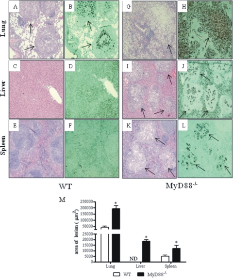
Fig. 4.
Photomicrographs of lesions of WT (A to F) and MyD88−/− (G to L) mice at week 8 of infection with 1 × 10⁶ P. brasiliensis yeast cells. Compared with those of MyD88−/− mice (G), the pulmonary lesions of WT mice (A) were smaller (arrows) and composed of organized granulomas containing lower numbers of yeast cells (B). The pulmonary lesions of MyD88−/− mice were composed of confluent, necrotic, unorganized granulomas of various sizes (G) containing an elevated number of fungal cells (arrow in H) and replaced almost all the normal tissue (G, H). The livers (C, D) and spleens (E, F) of WT mice presented a normal morphology; in contrast, the livers (I, J) and spleens (K, L) of MyD88−/− mice presented extensive necrotic lesions (arrows in I and K) containing an elevated number of yeast cells (arrows in panels J and L) surrounded by mononuclear inflammatory exudates. H&E (A, C, E, G, I, K)- and Groccot (B, D, F, H, J, L)-stained lesions (magnification, ×100). *, P < 0.05. (M) Total area of lesions in the lungs, livers and spleen of mice (n = 6) at week 8 after infection. *, P < 0.05.

