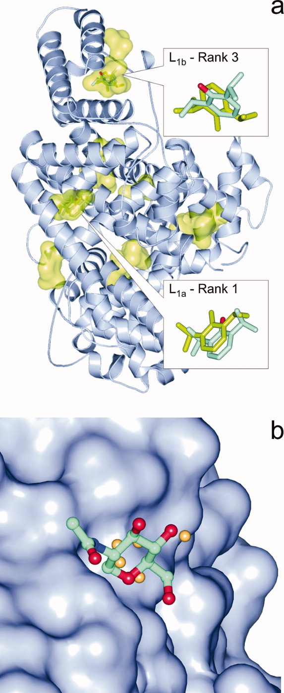
Figure 3.

(a) In PDB structure 1e7a two binding pockets of the primary ligand (L1) propofol had been detected by crystallography. Whereas the L1a pocket and the binding mode was identified precisely by BD (shown in inset as green sticks) as Rank 1 and Q-SiteFinder, the L1b pocket was located by PS methods and BD found it as Rank 3 (green sticks) with a rather high deviation from the crystallographic position (sticks colored by atom type). Other pockets found by BD are also shown as green surfaces. (b) Sitehound identified the shallow pocket (protein shown as surface) of NAG in the complex 1eqg-L2d in a 2.9 Å distance from the crystallographic ligand position (balls and sticks colored by atom type) by placing a few Carbon probes (beige spheres) into the proposed pocket. The small number of probes resulted in a low TIE value and a mis-ranking of this real binding pocket into the 88th of 112 ranks. [Color figure can be viewed in the online issue, which is available at wileyonlinelibrary.com.]
