
Fig. 2.
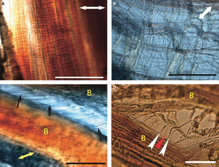
Images (A) and (B) taken simultaneously from adjacent regions under identical stress, demonstrating variation in ligament strain during constant stress in the ACL in fully hydrated, unfixed tissue, longitudinal sections. Double-headed white arrows indicate direction of applied stress. (A) Tightly adherent collagen bundles with no lateral separation with stress applied perpendicularly to collagen bundles. Scale bar: 100 μm (B) Interfascicular region demonstrating loose but organised tissue following application of stress perpendicular to ligament fascicles. Interfascicular fibres run obliquely to fascicles in both directions. Scale bar: 100 μm. (C) Variation in interbundle strain during application of perpendicular stress in the CLs in fully hydrated, unfixed tissue, longitudinal section, PCL × 40. Direction of applied strain is shown by yellow double-headed arrow. Collagen bundles are marked as yellow ‘B’. Straight, thick transverse interbundle fibres (black arrows) are seen. Scale bar: 100 μm (D) Variation in interbundle strain during application of perpendicular stress in the CLs in fully hydrated, unfixed tissue, longitudinal section, ACL × 40. Collagen bundles are marked as yellow ‘B’. Unilateral oblique interbundle fibres (white arrowheads). Subdivision of these oblique fibres is noted (red arrowhead). Scale bar: 40 μm.