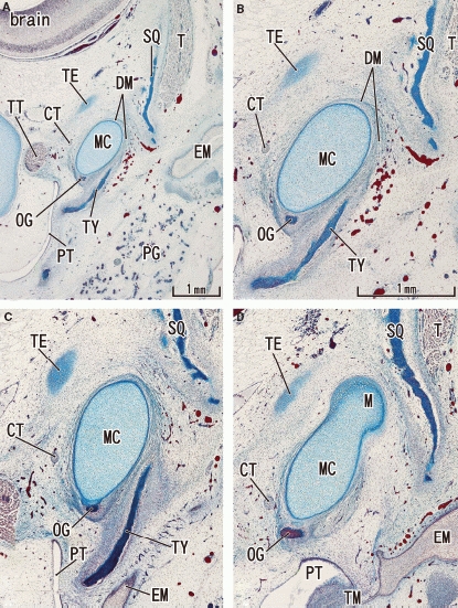
Fig. 2.

Tegmen tympani of the petrous part of the temporal bone appears in a 11-week human fetus. Frontal sections. Panel A (or D) most ventral (or dorsal) level of the figure. To show the topographical anatomy, panel A was prepared at lower magnification than panels B–D. The anlage of the tegmen tympani (TE) is a small oval cartilage located cranially and medially to the area of the tympanosquamosal fissure. Note the growth of the tympanic bone (TY) from the 10-week stage shown in Fig. 1. The squamous part (SQ) of the temporal bone narrows the tympanosquamosal fissure. Panels B–D are prepared at the same magnification (scale bar in panel B). CT, chorda tympani nerve; DM, discomalleolar ligament; EM, external auditory meatus; MC, Meckel's cartilage; OG, os goniale; PG, parotid gland; PT, pharyngotympanic tube; T, temporalis muscle; TM, tympanic membrane; TT, tensor tympani muscle and its tendon.
