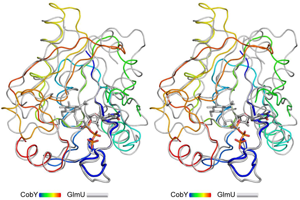
Figure 3. Structural comparison of CobY with the uridyltransferase domain of MtbGlmU.
MjCobY is depicted in a rainbow coloration scheme (N-terminus blue), whereas the uridyltransferase domain is colored in gray. The wide diameter ribbon indicates the consensus pyrophosphorylase motif. The structures are remarkably similar considering the lack of sequence similarity. Indeed, the rms difference between 144 structurally similar α-carbons is only 1.84 Å. The coordinates for MtbGlmU were obtained from the RCSB, accession number 3DJ4 (44). Several surface loops (151–176 and 197–222) were removed from GlmU to emphasize the similarity between these proteins.

