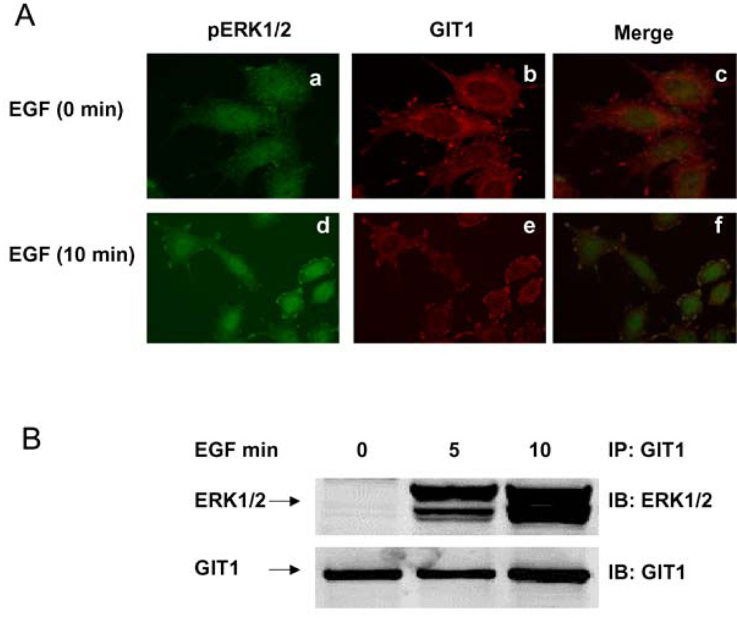
Figure 1. GIT1 associates with pERK1/2 in HeLa cells.
(A) HeLa cells were serum-starved for 6 h and stimulated with saline (a–c) or 10 ng/ml EGF (d–f) for 10 min. Cells were fixed with 4% formaldehyde and stained with an anti-pERK1/2 antibody (a and d) or an anti-GIT1 antibody (b and e). Panels c and fare the merged images. (B) HeLa cells were serum-starved for 6 h and stimulated with EGF (10 ng/ml) for the times indicated. Cytoskeleton fractions were immunoprecipitated with an anti-GIT1 antibody and probed with an anti-ERK1/2 antibody (top panel). To confirm equal protein loading, the blot was reprobed with the anti-GIT1 antibody (bottom panel). These results were reproducibly obtained in three independent experiments. IB, immunoblot; IP, immunoprecipitation.

