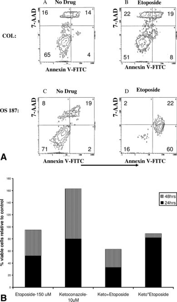
FIGURE 8.
(A) COL and OS187 cells were cultured with 150 µM etoposide for 24 hours and then examined by flow cytometry, using FITC-annexin V and 7-amino-actinomycin D (7AAD). Viable cells do not stain with either reagent. Staining with 7-AAD alone indicates necrotic cells. Staining with Annexin V identifies apoptotic cells and 7AAD staining separates early apoptotic (7AAD−) from late apoptotic (7AAD+) cells. (B) Viable COL cells remaining after exposure to etoposide, or ketoconazole and in combination for 24 hours (−) and 48 hours (−). (Keto+ represents simultaneous addition of both agents; Keto* represents 24 hours pretreatment with ketoconazole before the addition of etoposide.)

