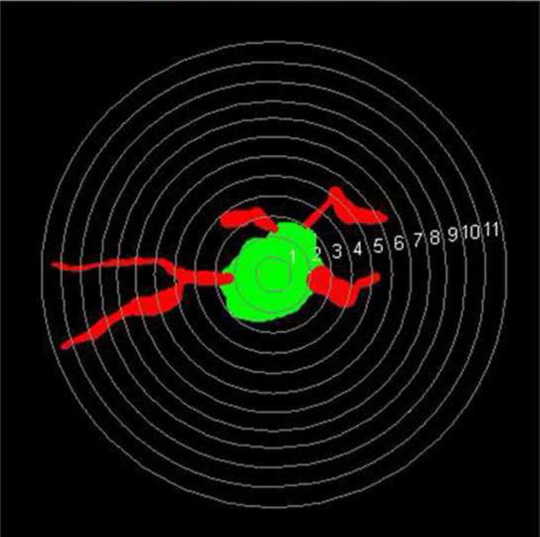
Figure 2.
A randomly selected parvalbumin-positive neuron with MAP2-positive dendrites selected from layer 6 from normal monkey S1 for dendritic complexity analysis is shown. Concentric spheres with different radii (1–11µm) with intervals of 0.5 µm are placed on the cell and its dendrites. The intersections between these circles and dendrites are used to analyze the complexity of dendritic arborization. (Sholl analysis, Neuroexplorer, Colchester, VT).

