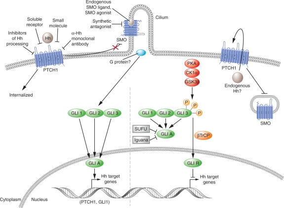
Figure 1.
Hedgehog signaling pathway in vertebrates. The above model illustrates our current understanding of the vertebrate Hedgehog (Hh) pathway signaling. Hh signaling cascade is initiated in the target cell by the Hh ligand binding to the Patched 1 protein (PTCH), a 12-span transmembrane protein located on the plasma membrane. Smoothened (SMO), a 7-transmembrane-span protein receptor, is located on the membrane of the intracellular endosome. In mammalians, the Hh signaling takes place in the nonmotile cilia to which the SMO and other downstream pathway components transit to in order to activate the GLI transcription factors [Rubin and de Sauvage, 2006; Corbit et al. 2005; Huangfu and Anderson, 2005; Huangfu et al. 2003]. An endogenous small molecule acting as a SMO agonist is transported outside the cell by PTCH, preventing its binding to SMO. In the absence of a Hh ligand, PTCH catalytically inhibits the activity of SMO by affecting its localization to the cell surface. Full-length GLI proteins are thus proteolytically processed to generate the repressor GLIR, largely derived from GLI 3, which represses Hh target genes. Binding of Hh to PTCH, internalizes and destabilizes PTCH, so that it can no longer transport the endogenous SMO agonist molecules outwards. Intracellular accumulation of this agonist molecule activates SMO which translocates to the plasma membrane, apparently concentrating in the cilia. Relief of SMO inhibition promotes generation of the activator GLIA, largely contributed by GLI 2 and the subsequent expression of the Hh target gene [Taipale et al. 2002]. CK1α, casein kinase 1α; GPCR, G-protein-coupled receptor; GSK3β, glycogen synthase kinase 3β; PKA, protein kinase A. Reprinted by permission from Macmillan Publishers Ltd: Rubin, L.L. and de Sauvage, F.J. (2006) Targeting the Hedgehog pathway in cancer. Nat Rev Drug Discov 5: 1026–1033.

