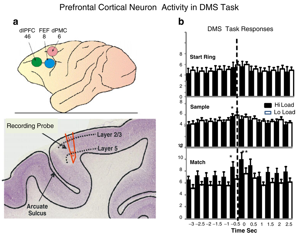
Fig. 2.
Prefrontal cortex (PFC) recording areas and neuron activity in DMS task. a Upper: areas 6, 8, and 46 of NHP prefrontal cortex which bracketed recording tracks for PFC neurons. Lower: cross section of NHP location of recording probe track near the arcuate sulcus in the premotor area. dlPFC dorsolateral prefrontal cortex, FEF frontal eye fields, dPMC dorsal premotor cortex. b Firing of PFC cells in each phase of the DMS task as a function of low vs. high cognitive load for juice reward trials. The PEHs show mean firing rate across all PFC cells (n=96) for the Start Ring Response, Sample Response, and Match Response at t=0.0 s (dotted line), on low (white bars) vs. high (black bars) cognitive load trials. Asterisks indicate significant differences in mean firing rate stated in text

