Fig. 1.
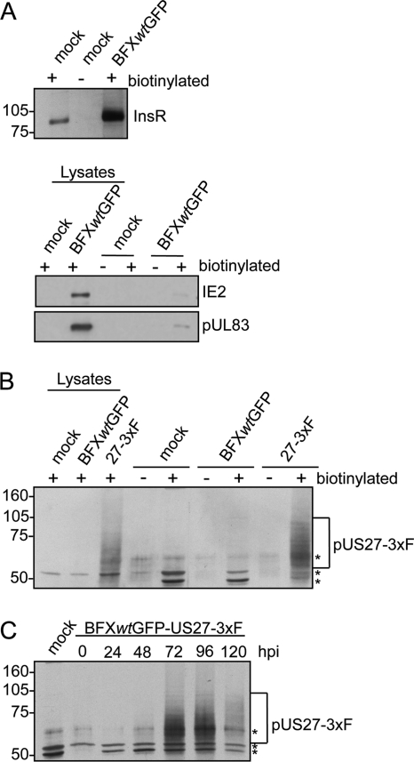
pUS27 is expressed on the surface of infected cells. Fibroblasts were mock infected or infected with BFXwtGFP or BFXwtGFP-US27-3×F at a multiplicity of 0.5 PFU/cell. Cell surface proteins were biotinylated 96 h later and captured with avidin-conjugated beads. Each lysate sample assayed received protein from 1.4 × 10⁵ cells per lane, and each biotinylated sample assayed was the material captured from 7.9 × 10⁵ cells. (A) Detection of cell surface insulin receptor InsR in biotinylated (+) samples but not in untreated cells (−) by immunoblot assay using InsR-specific antibody (top) and minimal detection of IE2 (nuclear localization) and pUL83 (nuclear plus cytoplasmic localization) in biotinylated samples (bottom). (B) Detection of FLAG-tagged pUS27-3×F in whole-cell lysates and in biotinylated samples by immunoblot assay using antibody to the FLAG epitope. The bracket marks glycosylated pUS27-3×F, and the asterisks denote nonspecific bands due to biotinylation. (C) Peak expression of pUS27-3×F at 72 and 96 hpi. Fibroblasts were biotinylated at various times after infection and then assayed as in panel B.

