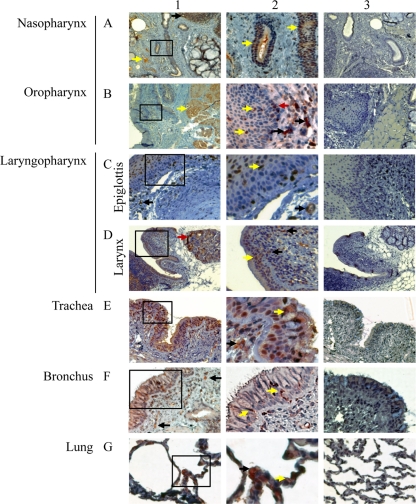
Fig. 2.
Detection of ACE2+ cells by immunohistochemistry at multiple levels of the respiratory tracts of rhesus macaques. Positive labeling for ACE2 was found at all levels of the respiratory tract. For each level examined, a low-magnification overview is shown in panel 1 with a higher magnification of the boxed area in panel 1 shown in panel 2. Panel 3 shows the same tissue immunolabeled with control rabbit serum. The magnifications of each image are as follows: ×40 (A1), ×100 (A3, B1, B3, C1, C3, D1, D3, E1, and E3), ×200 (A2, F1, F3, G1, and G3), and ×400 (B2, C2, D2, E2, F2, and G2). In nasopharynx (A) and oropharynx (B), ACE2+ cells were identified in lymphatic nodules (black arrow, A1), vascular endothelium (yellow arrow, A1), the epithelium of salivary gland ducts (yellow arrow, A2), some muscle cells (yellow arrow, B1), epithelium lining (yellow arrows, B2), lamina propria (black arrows, B2), and basal layer of the epidermis (red arrow, B2). In epiglottis (C), ACE2+ cells were found in lamina propria (black arrow, panel 1), the epithelial lining (yellow arrow, panel 2), and capillaries (black arrow, panel 2). In larynx (D), ACE2+ cells were observed in glands (red arrow, panel 1), the epithelial lining (yellow arrow, panel 2), and lamina propria (black arrows, panel 2). In trachea (E), numerous ACE2+ cells were found in the epithelial lining (yellow arrow, panel 2), and lamina propria (black arrow, panel 2). In bronchus (F), many ACE2+ cells were identified in lamina propria (black arrows, panel 1) and in the epithelial lining (yellow arrows, panel 2). In lungs (G), ACE2+ cells were observed on type I pneumocytes (yellow arrow, panel 2) and type II pneumocytes (black arrow, panel 2).

