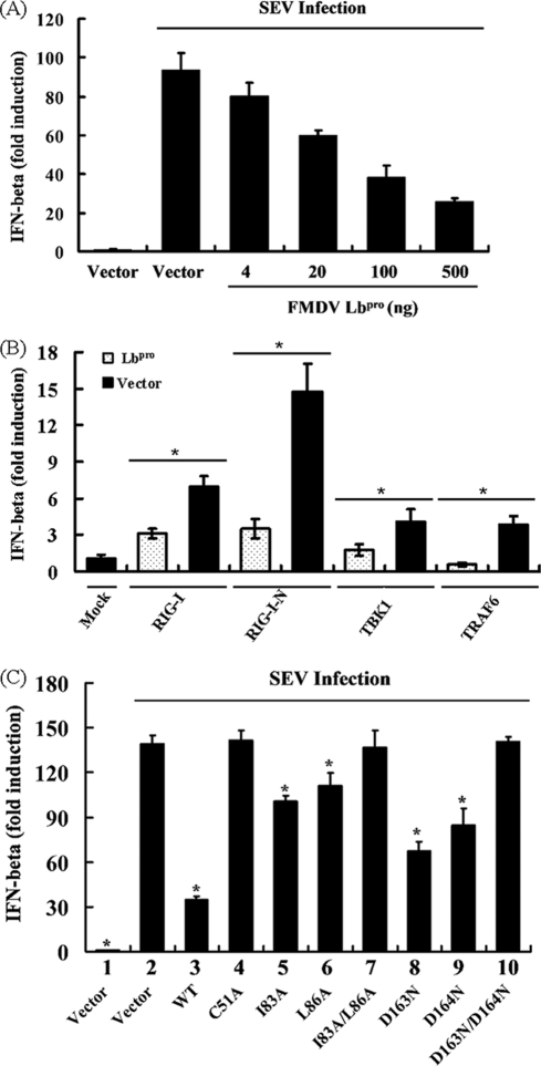
Fig. 6.
Lbpro inhibits type I interferon induction. (A) HEK293T cells grown in 24-well plates were transfected with 0.1 μg/well of IFN-β-Luc reporter plasmid, along with 0.05 μg/well of pRL-TK plasmid and increasing quantities (0, 0.004, 0.02, 0.1, or 0.5 μg) of plasmid encoding Lbpro, using Lipofectamine 2000. Twenty-four hours after the initial transfection, the cells were further infected with SEV or mock infected. Luciferase assays were performed at 18 h after infection. Results represent the means and standard deviations from three independent experiments. The relative firefly luciferase activity was normalized to the Renilla reniformis luciferase, and the untreated empty-vector control value was set to 1. (B) HEK293T cells were cotransfected with the IFN-β-Luc reporter plasmid (0.1 μg), pRL-TK plasmid (0.05 μg), and 0.5 μg of plasmid encoding Lbpro together with the RIG-I, RIG-I N, TBK1, or TRAF6 expression vector (0.5 μg). Luciferase assays were performed at 36 h after transfection. (C) HEK293T cells were cotransfected with the IFN-β-Luc reporter plasmid (0.1 μg), 0.05 μg of pRL-TK, and the designated Lbpro expression plasmids (0.5 μg). An empty vector (pcDNA3.1-V5/His B) was used as a control. Twenty-four hours after the initial transfection, the cells were further infected with SEV or mock infected. Cell extracts were collected at 18 h after infection and analyzed for firefly and Renilla luciferase expression. *, P < 0.01 compared with vector plus SEV.

