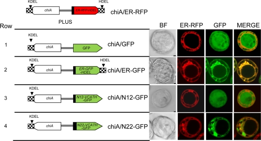
Fig. 3.
Fluorescence patterns of V-CATH-GFP fusion proteins from coinfection of Hi5 cells with chiA/ER-RFP (top) and the constructs shown on the left. Bright-field (BF) and fluorescence patterns of virus-expressed V-CATH-GFP fusion proteins (chiA/N12-GFP or chiA/N22-GFP [rows 3 and 4]) were compared to those of the diffuse GFP control virus (chiA/GFP [row 1]) or the ER-targeted GFP control virus (chiA/ER-GFP [row 2]) in infected Hi5 cells. In all rows, the ER was labeled with red fluorescence by coinfection with the chiA/ER-RFP virus. For N12-GFP and N22-GFP, the first 12 or 22 amino acids, respectively, encoded by the v-cath ORF are fused to the amino terminus of GFP. Hi5 cells were infected at an MOI of 10. Virus-infected cells were photographed at 40 hpi using confocal laser scanning microscopy.

