Fig. 3.
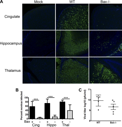
Bax is important for reovirus growth in the brain. WT and Bax−/− mice were injected with 100 PFU T3A i.c. and sacrificed at 8 dpi. (A) Coronal sections were stained by immunofluorescence for reovirus antigen σ3 (green) and Hoechst (blue) in the cingulate cortex (Cing), hippocampus (Hippo), and thalamus (Thal) of mock- or T3A-infected WT and Bax−/− mice (100×). An asterisk indicates the antigen-positive ependymal cells lining the lateral ventricle in the Bax−/− brain. (B) The percentage of reovirus-infected neurons was determined per high-power field (HPF) (400×) in the cingulate (n = 14 for WT [Bax+], and n = 10 for Bax−/− [Bax−]), hippocampus (n = 12 for WT, and n = 10 for Bax−/−), and thalamus (n = 14 for WT, and n = 9 for Bax−/−). Lines between bars indicate comparisons. ***, P < 0.0001. (C) Viral titers from WT (closed circles) and Bax−/− (open circles) brains of mice infected with 100 PFU T3A at 8 dpi. Each circle represents an average viral titer from one mouse brain.

