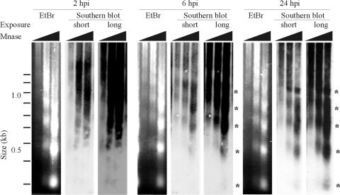
Fig. 3.
hdAd vector DNA assembles into physiologically spaced chromatin after host cell transduction. A549 cells were infected with hdAd-PGK-mSEAP (MOI = 500), and the samples were processed for micrococcal nuclease (MNase) assay as described in Materials and Methods. The ethidium bromide (EtBr)-stained agarose gel (showing bulk cellular chromatin) and short and long autoradiograph exposures (showing Ad DNA) are displayed. Bands representing DNA that was protected from MNase cleavage are indicated by asterisks.

