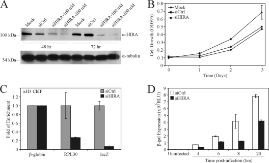
Fig. 5.
Deposition of H3.3 promotes efficient expression of virus-carried genes. (A) HeLa cells were transfected with control siRNA (siCtrl) or siRNA targeting HIRA mRNA (siHIRA), and 48 or 72 h posttransfection, the cells were analyzed by immunoblotting for expression of HIRA or tubulin. (B) HeLa cells were transfected with 100 nM siCtrl or siHIRA and, 48 h later, replated to evaluate cellular growth kinetics. OD595, optical density at 595 nm. (C) HeLa cells were transfected with siCtrl or siHIRA, and 48 h later, the cells were infected with hdAd-lacZ (MOI = 10). Six hours later, the cells were processed for ChIP with anti-H3 or IgG antibody. The resulting ChIP DNA was analyzed by qPCR for the presence of hdAd DNA (lacZ amplicon), the cellular β-globin, or RPL30 loci. The level of enrichment with H3 is expressed relative to that of siCtrl (n = 4). (D) HeLa cells were transfected with siCtrl or siHIRA, infected 48 later with hdAd-lacZ (MOI = 10), and analyzed for β-Gal activity at various time postinfection (n = 3; means ± standard errors of the means).

