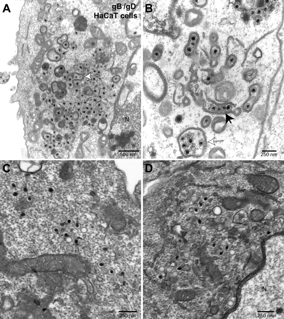
Fig. 9.
Electron microscopy of HaCaT cells infected with F-BAC gB−/gD−. HaCaT cells were infected with F-BAC gB−/gD− for 24 to 28 h and then fixed, stained, and sectioned for electron microscopy. The filled arrow in panel B points to a row of partially enveloped capsids, i.e., partially wrapped with a double membrane. Open arrows in panels A and B point to multiple capsids apparently fully wrapped in a double membrane. N, nucleus.

