Fig. 2.
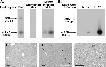
Vertical transmission of MCMV following acute infection. (A) Lactating mice were infected (i.p.) with 300 PFU of MCMV-GFP at 6 days after birth of the litter. At 11 days of lactation, breast milk leukocytes were collected from an uninfected control mother or from two infected lactating mothers (5 days after virus infection). Total RNA was examined for MCMV IE-1 gene expression by RT-PCR with Southern analysis. The IE-1 primers bracket an intron, allowing a distinction between spliced viral mRNA (188 bp) and contaminating genomic DNA (310 bp) (confirmed by analysis of total RNA extracted from peritoneal cavity [PerC] exudate cells without DNase treatment from an acutely infected mouse). (B) At 5, 7, 9, or 12 days after maternal infection, nursing pups were sacrificed and lungs from individual neonatal mice were examined for expression of MCMV IE-1 mRNA by RT-PCR with Southern analysis. (C to E) Histological evaluation of liver sections from neonates nursed for 1 week by an uninfected mother (C) or an acutely infected mother (D and E). Sections were stained with hematoxylin and eosin. Arrows indicate inflammatory foci (D); an enlargement of the marked area is shown in panel E. Scale bar, 50 μm.

