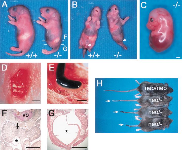
Figure 2.
Caudal truncation in CYP26 mutant mice. (A) Wild-type (+/+) and CYP26−/− mice at postnatal day (P) 1, with the mutant exhibiting sirenomelia. The levels of the sections shown in F and G are indicated. (B) Ventral view of wild-type and CYP26−/− mice at P1. The mutant exhibits sirenomelia and lacks external genitalia (arrowhead). (C) Lateral view of an E16.5 CYP26−/− embryo completely lacking hind limbs and a tail. (D) Dorsal view of a homozygous mutant embryo at P1 exhibiting spina bifida. (E) Atresia of the gut in a CYP26−/− embryo at P1. (F,G) Histological examination of the abdomen of a CYP26−/− mouse at P1. Horseshoe kidney (arrow) and a fused ureter (asterisk) are apparent in F. The abnormally dilated bladder (asterisk) apparent in G connects to the ureter shown in F. (vb) Vertebra body. (H) CYP26neo/neo and CYP26neo/− mice at 8–10 wk after birth. Various types of kinky tail (arrows) are apparent in the CYP26neo/− animals. Scale bar, 1 mm for C–G.

