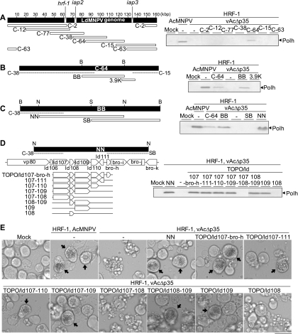
Fig. 1.
Identification of an LdMNPV gene that is responsible for the suppression of apoptosis in Ld652Y cells infected with vAcΔp35. (A) Seven cosmid clones (C-2, C-12, C-77, C-38, C-64, C-15, and C-63), which cover the entire LdMNPV genome (left), were each transfected into Ld652Y cells along with the plasmid pHyHr6IE1/HA-HRF-1 harboring the hrf-1 gene. Twenty-four hours after transfection, the cells were infected with vAcΔp35 at a multiplicity of infection of 0.1. The transfected and infected cells were analyzed by immunoblot analysis with antipolyhedrin antibody for polyhedrin (Polh) production at 72 h postinfection (right). The polyhedrin signals were visualized by using horseradish peroxidase (HRP)-conjugated goat anti-rabbit IgG antibody (Zymed) and a Konica immunostaining HRP-1000 kit (Konica). An LdMNPV genomic map with scales of kilobase pairs (kbp) is presented, together with the locations of the hrf-1, iap2, and iap3 genes. (B) The BB fragment, which was derived from cosmid C-64 through digestion with BamHI, and the PCR-amplified 3.9K DNA fragment (left) were cloned into pBlueScript and transfected into Ld652Y cells together with hrf-1. The transfection-infection experiments were performed as described for panel A for analysis of polyhedrin production (right). (C) The NN and SB fragments, which were derived from the BB fragment by digestion with NotI or SmaI (left), were similarly tested for polyhedrin production (right). (D) Eight expression plasmids harboring one or multiple gene(s) carried on the NN fragment (TOPO/ld107-bro-h, TOPO/ld107-111, TOPO/ld107-110, TOPO/ld107-109, TOPO/ld107-108, TOPO/ld108-109, TOPO/ld109, and TOPO/ld108) (left) were similarly tested for polyhedrin production (right). (E) Images obtained by microscopy showing vAcΔp35-infected Ld652Y cells previously cotransfected with the plasmid pHyHr6IE1/HA-HRF-1 harboring the hrf-1 gene and each of the expression plasmids containing subsets of the six putative viral genes carried by the NN fragment, presented in the left side of panel D. The arrows indicate the cells containing polyhedra. The scale bar indicates 30 μm. Restriction endonuclease sites in panels B to D are as follows: B, BamHI; N, NotI; S, SmaI.

