Fig. 2.
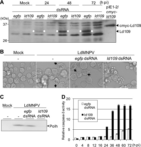
Effects of RNAi-mediated silencing of ld109 on the induction of apoptosis in Ld652Y cells infected with LdMNPV. (A) RNAi silencing of Ld109 protein. Monolayer cultures of 1 × 10⁶ Ld652Y cells in 35-mm culture dishes were transfected with 1 μg of dsRNAs against ld109 and the EGFP gene. At 24 h posttransfection, the cells were infected with LdMNPV at a multiplicity of infection of 1. At 24, 48, and 72 h postinfection (h pi), these cells were examined for the expression of Ld109 by immunoblot analysis with anti-Ld109 antibody as the primary antibody and HRP-conjugated goat anti-rabbit IgG antibody as the secondary antibody. Polypeptides from LdMNPV-infected Ld652Y cells were resolved on a 12.5% SDS–polyacrylamide gel and blotted onto an Immobilon-P transfer membrane (Millipore). The Ld109 protein signals were visualized by enhanced chemiluminescence Western blotting detection reagents (Amersham Biosciences) and are highlighted by the white dots. The mock-infected cells previously transfected with dsRNAs against ld109 and the EGFP gene and the cells transfected with pIE1-2/cmyc-ld109 expressing cMyc-Ld109 protein were also analyzed. BenchMark prestained protein ladder (Invitrogen) was used as the protein size marker. (B) Images obtained by microscopy show the apoptosis induced by RNAi silencing of ld109 in LdMNPV-infected Ld652Y cells. Cells were observed at 96 h posttransfection (72 h postinfection) with dsRNAs against ld109 and the EGFP gene. The arrows indicate the cells containing polyhedra, and the scale bar indicates 30 μm. (C) Immunoblot analysis of polyhedrin (Polh) production. The transfected and infected cells were harvested at 72 h postinfection and analyzed by using antipolyhedrin antibody as the primary antibody and HRP-conjugated goat anti-rabbit IgG antibody as the secondary antibody. Polypeptides were resolved on a 12.5% SDS–polyacrylamide gel and blotted on a nitrocellulose membrane (Advantec Toyo). The polyhedrin signals were visualized by using a Konica immunostaining HRP-1000 kit. (D) Caspase-3-like protease activity of the cells transfected with ld109 dsRNA. The LdMNPV-infected cells previously transfected with dsRNAs against ld109 and the EGFP gene were harvested at the indicated times postinfection, and the caspase-3-like protease activities of these cells were determined by using Ac-DEVD-AMC (N-acetyl-Asp-Glu-Val-Asp-7-amino-4-methylcoumarin) as the substrate. The caspase-3-like protease activities are presented as the ratios to the activity of mock-infected cells at 0 h postinfection. Vertical bars indicate standard deviations of averages from three determinations.

