Fig. 1.
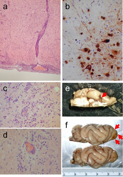
Brain lesions in HK486 virus-infected ferrets. (a) Severe nonsuppurative encephalitis in the olfactory area at day 6 postinfection (p.i.). (b) Viral antigen expression in a brain lesion at day 12 p.i. Neuronal and glial cells are stained with anti-H5 virus antiserum. (Inset) Noninfected neurons and glia. (c) Smoldering encephalitis in brain tissue at 3 months p.i. (d) Perivascular glial scar formation at 9 months p.i. (e) Macroscopically visible brain lesion in part of the olfactory system (piriform lobe) on day 12 p.i. (f) Partial loss of an olfactory bulb (upper side of brain [red arrows]) due to viral encephalitis at 1 month p.i. Compare these images with the brain from an age-matched control (lower brain).

