Fig. 2.
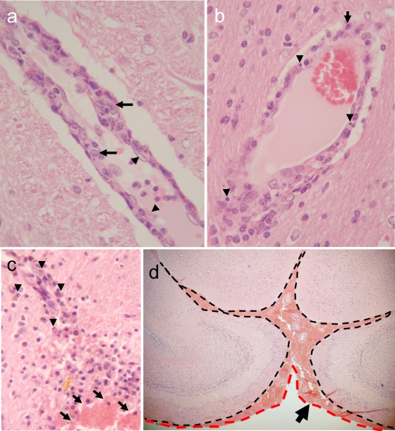
Brain lesions in HK483 virus-infected ferrets. (a) Prominent nonsuppurative vasculitis at day 6 p.i. Note the severe swelling of a vascular endothelial cell (arrowheads) and migration of macrophages into the vascular wall (arrows), compared with the normal appearance of the surrounding brain parenchyma. (b) Scattered apoptotic cells (arrowheads) and polymorphonuclear leukocytes (arrow) in the vascular wall on day 6 p.i. (c) Old and fresh hemorrhagic lesions in the thalamus of a ferret that underwent necropsy at 6 months p.i. (arrowheads, hemosiderin-laden macrophages in an old lesion; arrows, fresh hemorrhage with red blood cells). (d) Fresh subarachnoid hemorrhage in a ferret brain at 6 months p.i. Note the accumulation of red blood cells between leptomeninges (dashed black line) and arachnoid mater (dashed red line).

