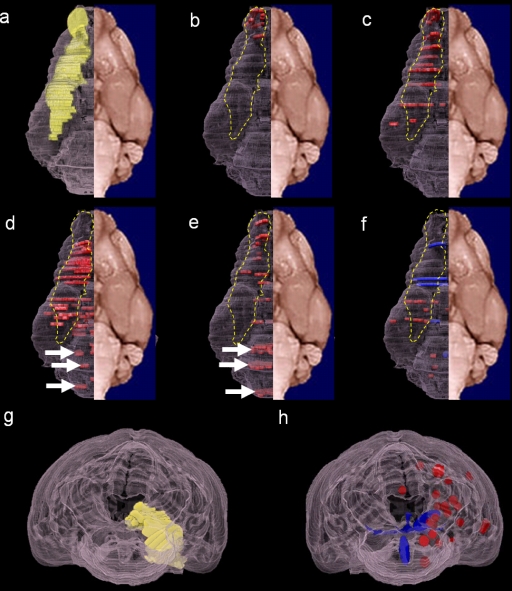
Fig. 3.
Distribution of brain lesions following infection with the HK483, HK486, HK213, NCVD18, or VN1204 strain of H5N1 virus. Brain lesions are mapped on three-dimensional images of a yellow mongoose brain. Selected parts of the brain sections were analyzed; therefore, the plots of the lesion locations are discontinuous. (a) Olfactory route (yellow). The distribution of lesions and viral antigens (red) associated with HK213 (b) or NCVD18 (c) infection follows the olfactory route (dashed yellow line). In animals infected with HK486 (d) or VN1204 (e), the lesions and viral antigens are located in the brain stem (white arrows) and the olfactory route (red plots within the dashed yellow line). The HK483 strain (f) caused severe blood vessel damage, with apparent hemorrhagic lesions (blue plots). (g) Posterior view of the olfactory route (yellow plots). (h) Posterior view of HK483-induced hemorrhagic lesions (blue) and vasculitis (red) outside the olfactory route. Panels a to f are ventral views.

