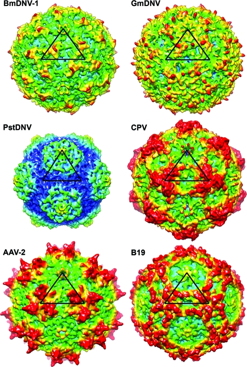
Fig. 4.
Comparison of the BmDNV-1 protein capsid with those of other parvoviruses. Surface renderings of three-dimensional electron density maps of BmDNV-1, GmDNV, PstDNV, CPV, B19, and AAV-2 generated from atomic coordinates to 8-Å resolution. One icosahedral asymmetric unit is indicated by a black triangle. The surface is colored according to the distance from the viral center (blue, 100 Å; cyan, 107.5 Å; green, 115 Å; yellow, 122.5 Å; red, 130 Å).

