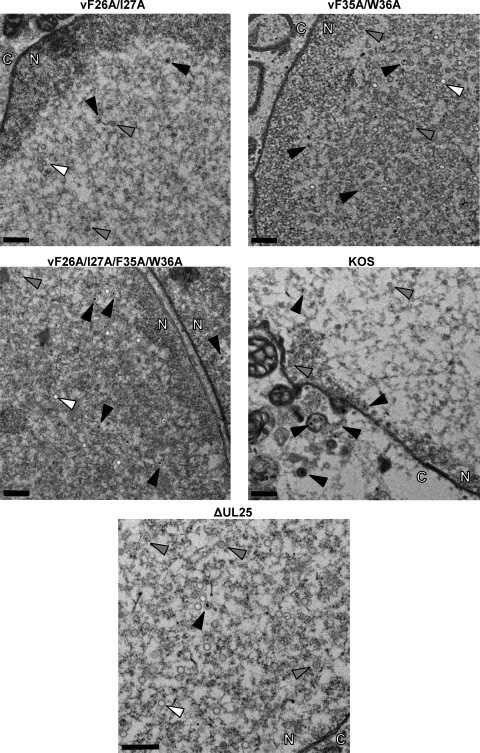
Fig. 5.
Transmission electron micrographs of thin-section preparations of virus-infected cells. Vero cells were infected with the indicated viruses at an MOI of 5, and at 18 hpi the cells were fixed and sectioned for imaging. The arrows point to A capsids (white arrows), B capsids (gray arrows), and C capsids (black arrows). The nucleus and cytoplasm are marked with N and C, respectively. Magnification of top four panels, ×4,400; magnification of bottom panel, ×6.500. Scale bars, 0.5 μm.

