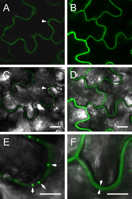
Fig. 7.
Subcellular localization of GFP-MP versus GFP in N. benthamiana leaf epidermal cells, observed 3 days postagroinfiltration. Images show localization of pGC-GFP-MP (A, C, and E) and pGC-RNA3-GFP (B, D, and F) in agroinfiltrated leaves. The fluorescence images in panels A and B are overlaid with the corresponding bright-field images in panels C and D; only the overlaid pictures are shown for the higher magnification images in panels E and F. In panels A and C the punctate pattern of MP-GFP fluorescence and its association with the cell wall are visible (arrowhead); panel E shows a particularly evident pattern (arrowheads). Localization of free GFP (B and D) to the cytoplasm clearly identifies the unlabeled cell wall (F) as a dark line separating the fluorescent cytoplasms of adjacent cells (arrowheads). Bars, 10 μm (A to D) and 5 μm (E and F).

