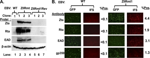
Fig. 3.
Spontaneous reactivation of EBV into lytic replication in 293-D cells latently infected with ZIIRmt, but not WT virus. (A) Immunoblots showing relative levels of EBV Zta, Rta, and EAD proteins present in 293-WT, 293-ZIIRmt(Rm), and 293-ZIIRmtRev cell lines. The cellular protein β-actin present in the same samples served as a loading control. (B) IFS of 293-WT1 and 293-ZIIRmt(Rm clone1) cells for the presence of Zta, Rta, EAD, and gp350 proteins. The primary antibodies used are indicated to the left of each row of images. Fields of cells were photographed with different filters to show the total EBV-positive ones (GFP) versus the subset of them containing the indicated EBV-encoded protein (IFS). The percentages of IFS-positive cells are indicated to the right of each field; these values were obtained by scoring 10 fields of cells.

